Granulation Tissue Explained (2026 Guide): What It Is, How Wounds Heal & Warning Signs
That pink, bumpy tissue in a healing wound has a name - granulation tissue. Here is what it does, what healthy healing looks like, and the signs that need a clinician's eye.
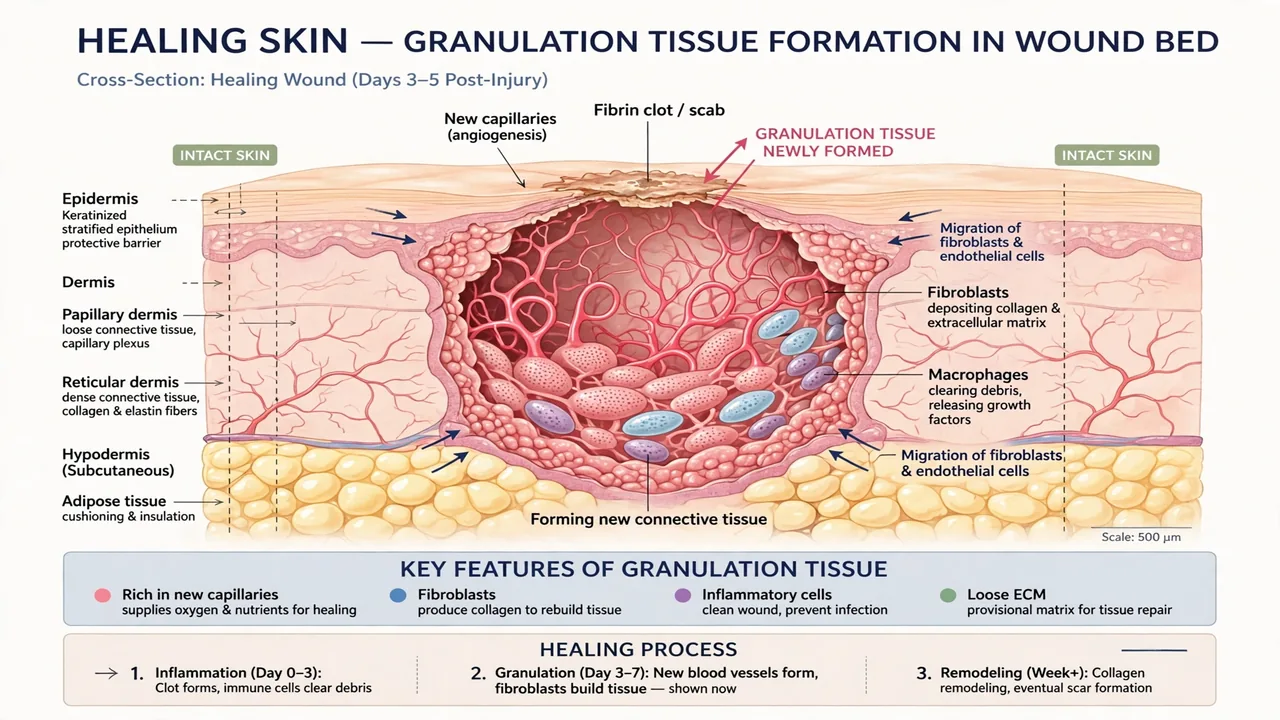
If you have ever watched a scraped knee or a surgical incision heal, you have seen granulation tissue – even if you did not know its name. It is the pinkish, slightly bumpy tissue that fills in the wound bed a few days after an injury, looking a little like tiny red cobblestones. People often notice it and worry: Is this normal? Is it infected? Should I leave it alone? This guide answers those questions in plain language: what granulation tissue is, what it does, what healthy healing looks like, and which signs mean it is time to call a clinician.
A quick note before we start: this article is general information, not medical advice. Wounds that are deep, large, not improving, or showing signs of infection deserve professional care – this guide will help you recognize those situations, not replace them.
In this article
- What granulation tissue is, in plain language
- The four phases of wound healing
- What healthy granulation tissue looks like
- Unhealthy granulation tissue: warning signs
- Hypergranulation (“proud flesh”)
- What slows granulation down
- How wounds are managed during this stage
- What you can do at home
- FAQs
What Is Granulation Tissue?
The short answer
Granulation tissue is new connective tissue that forms on the surface of a wound while it heals. It is built from three main ingredients: new capillaries (tiny blood vessels), fibroblasts (cells that produce collagen and other structural proteins), and the extracellular matrix they lay down – the scaffolding that holds everything together. The name comes from its granular, bumpy appearance: those bumps are the visible tops of new capillary loops growing upward into the wound bed.
What it is: New, temporary tissue – a mix of blood vessels, collagen-producing cells, and matrix – that fills a wound from the bottom up.
Where it appears: In the wound bed of open wounds healing by secondary intention – cuts, abrasions, surgical wounds left open, ulcers, and burns.
What it looks like: Pink to red, moist, and granular or bumpy, often described as resembling cobblestones.
When it forms: Typically beginning around day 3 to 5 after injury, during the proliferative phase of healing.
What it becomes: Over weeks to months it is remodeled into stronger, paler scar tissue.
Think of granulation tissue as the construction site of healing. Before the skin can close over a wound, the body has to rebuild the missing foundation – and granulation tissue is that foundation going up, beam by beam and vessel by vessel.
The Four Phases of Wound Healing
Granulation tissue does not appear out of nowhere. It arrives during the third of four overlapping phases that every healing wound moves through. Understanding the sequence makes the whole process less mysterious:
1. Hemostasis – stopping the bleeding
Within seconds of injury, blood vessels constrict and platelets form a clot. That clot is not just a plug; it is also a temporary scaffold and a reservoir of signaling molecules that summon the next wave of cells.
2. Inflammatory phase – cleaning up
Over the next few days, white blood cells flood the area to clear bacteria, debris, and damaged tissue. Redness, warmth, swelling, and some pain here are normal – this controlled inflammation is the cleanup crew at work. Problems arise only when inflammation drags on too long.
3. Proliferative phase – rebuilding
Starting around day 3 to 5, fibroblasts migrate in and begin producing collagen, new capillaries sprout into the wound (a process called angiogenesis), and granulation tissue fills the defect from the bottom up. Meanwhile, epithelial cells crawl in from the wound edges to resurface it. This is granulation tissue’s home phase.
4. Remodeling phase – strengthening
Over weeks to months, the hastily built collagen is reorganized into stronger, more orderly fibers. The wound contracts, the redness fades, and granulation tissue matures into scar tissue – which typically reaches only about 80 percent of the original skin’s strength, even in the best case.
These phases overlap rather than clicking from one to the next like gears. A wound can be finishing inflammation at its edges while proliferation is already underway in the center – which is why healing looks uneven and sometimes confusing from the outside.

What Healthy Granulation Tissue Looks Like
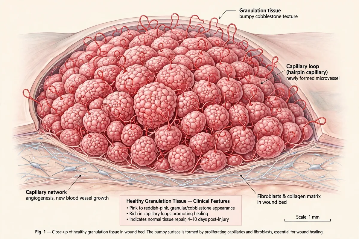
Healthy granulation tissue has a fairly distinctive look once you know what to check for:
- Color: pink to bright red, sometimes described as coral red – the color of good blood supply.
- Texture: granular and bumpy, like cobblestones – those bumps are new capillary loops.
- Moisture: moist and slightly glossy, not dry and not swimming in fluid.
- Growth pattern: fills the wound bed evenly and gradually, rising from the base upward.
- Sensitivity: usually not very painful to light touch; it may bleed a little if bumped because it is so vascular – light bleeding on contact alone is not automatically a problem.
If the tissue in a healing wound matches that description and the wound is steadily getting smaller, healing is on track. Steady progress is the single most reassuring sign in wound care – a wound that looks a little odd but shrinks week by week is usually fine, while a wound that looks “okay” but never changes deserves attention.
Unhealthy Granulation Tissue: Warning Signs
Granulation tissue can also signal trouble. The differences are worth learning because they are visible to anyone paying attention:
| Healthy sign | Warning sign | What it may mean |
|---|---|---|
| Pink to bright red | Dark, dusky, or purplish red | Poor blood supply or possible infection |
| Moist and glossy | Dry, or excessively wet and soggy | Moisture imbalance stalling healing |
| Bumpy, cobblestone texture | Smooth, flat, or pale | Weak granulation, poor perfusion |
| Minimal bleeding on light touch | Bleeds easily and excessively | Fragile tissue, possible infection or irritation |
| Steadily fills the wound | No new growth for a week or more | Stalled healing – needs evaluation |
| Little or no odor | Foul smell, pus, or cloudy drainage | Infection until proven otherwise |
A few specific situations deserve emphasis. Dark red tissue that bleeds at the slightest contact and is painful is the classic picture of unhealthy granulation tissue and should be seen by a clinician – infection or another underlying problem is often behind it. Pale, shiny, or white-ish tissue can indicate poor blood flow, while a yellow or white fibrous film over the wound is often slough – dead tissue that blocks healing and usually needs professional removal (debridement). And spreading redness, increasing pain, swelling, warmth, fever, or red streaks moving away from the wound are infection red flags that should not wait.
Hypergranulation: When There Is Too Much of a Good Thing
Sometimes granulation tissue overdoes it. Instead of stopping at the level of the surrounding skin, it keeps growing and rises above the wound surface as a red, fleshy, sometimes shiny mound. Clinicians call this hypergranulation – the older term “proud flesh” is still used, especially in veterinary wound care.
Overgrowth matters because skin cells cannot easily migrate across a raised mound to close the wound, so healing stalls. Common contributors include prolonged inflammation, excess moisture, friction from dressings or clothing, and low-grade infection or bacterial colonization. Management is a clinician’s job and typically starts with removing the trigger: controlling moisture, changing the dressing type, and treating any infection. Persistent overgrowth may be reduced with silver nitrate cautery or trimmed back surgically – quick office procedures, not home projects.
What Slows Granulation Down
When granulation tissue fails to appear or stalls, there is usually a reason. The most common ones:
- Infection. Bacteria prolong inflammation and damage new tissue; some form biofilms that resist both the immune system and antibiotics.
- Poor blood supply. New tissue needs oxygen and nutrients. Peripheral artery disease, pressure on the wound, and smoking all reduce perfusion.
- Diabetes. High blood sugar impairs white blood cell function, circulation, and collagen production – which is why diabetic foot ulcers are so prone to stalled healing.
- Moisture imbalance. Both extremes hurt: a dried-out wound bed kills new cells, while a constantly soggy one macerates tissue and invites infection. Modern wound care aims for a balanced moist environment.
- Repeated trauma. Picking at the wound, friction from clothing, or pressure on the area tears down new tissue as fast as the body builds it.
- Poor nutrition. Protein, vitamin C, and zinc all play documented roles in collagen synthesis and immune function; deficiencies slow repair.
- Certain medications. Corticosteroids and some immunosuppressants blunt the inflammatory and proliferative phases.
The practical takeaway: if a wound shows no new pink tissue after about two weeks, or if progress stalls for more than a week, that is the point to seek professional evaluation rather than waiting it out.
How Wounds Are Managed During This Stage
Clinicians managing a granulating wound are essentially protecting the construction site. The principles are consistent across settings:
Keep it clean, keep it covered, keep it balanced. Gentle cleansing, an appropriate dressing that maintains moist – not wet – conditions, and regular dressing changes matched to how much fluid the wound produces. Dressings are chosen for the wound: a dry, lightly draining wound gets something different from a heavily draining one.
Remove what does not belong. Dead tissue (slough or eschar) and excess hypergranulation are removed by debridement – which can be as gentle as dressings that encourage the body’s own enzymes, or as direct as a clinician trimming tissue with instruments.
Address the cause. Offloading pressure from a foot ulcer, improving blood sugar control, treating infection based on culture results, correcting nutrition – the wound is often the symptom and the underlying condition is the disease.
Protect the edges. The surrounding skin matters too; macerated or damaged wound edges cannot support the epithelial cells trying to close the wound.
What You Can Do at Home
For minor wounds healing normally, the home playbook is refreshingly boring – which is exactly the point:
- Leave the wound covered and change dressings as directed; uncovering it “to air out” dries the wound bed and slows healing.
- Wash your hands before touching the wound or dressing.
- Do not pick at scabs or new tissue – you are demolishing what the body just built.
- Eat normally and well; healing burns extra energy and protein.
- Take dated photos every few days. It is genuinely hard to judge slow change from memory, and photos give a clinician something concrete if you end up seeking care.
- Watch for the warning signs in the table above – especially spreading redness, increasing pain, pus, foul odor, fever, or red streaks – and get care promptly if they appear.
Recovery from injuries is a wider topic than wound beds alone – for example, this guide to practical ways to ease achy knees covers the day-to-day side of getting an injured joint comfortable again while the body does its repair work.
Frequently Asked Questions
Is granulation tissue a good sign?
Yes – in most cases it means the wound has moved past the inflammatory phase and is actively rebuilding. Pink, bumpy, moist tissue that gradually fills the wound is exactly what healing is supposed to look like.
Does granulation tissue hurt?
It is usually not very painful to light touch, though the wound overall may still be tender. New, sharp, or increasing pain – especially with redness, swelling, or discharge – is different and worth getting checked.
How long does granulation take?
It typically begins 3 to 5 days after injury and continues for days to weeks depending on the wound’s size and depth. Large or complicated wounds can granulate for much longer; small scrapes may barely show the phase at all.
What does white or yellow tissue in a wound mean?
White or yellow fibrous material is often slough – dead tissue that needs to be cleared before healing can proceed – rather than granulation tissue. It is one of the findings clinicians look for when a wound stalls, so mention it at your appointment rather than trying to remove it yourself.
Should I remove granulation tissue?
No. Healthy granulation tissue should be left alone – it is the wound healing itself. Only clinicians should remove tissue, and only when it is dead tissue or problematic overgrowth. Picking at a healing wound sets healing back.
Can granulation tissue turn into a scar?
Essentially, yes. During the remodeling phase, granulation tissue’s loose collagen is reorganized into denser, stronger fibers and the area becomes scar tissue – paler, flatter, and less elastic than the original skin.
Reead Also: Water Tower Water Tower: How These Giant Tanks Work (2026 Guide)
#granulationtissue #woundhealing #healthandwellness

Related Articles

Whippets: The Gentle Speedster Dog Breed (2026 Guide)
Whippets combine sprint speed with a calm, affectionate home life. This guide covers what the breed…
Canker Sore Treatment: 12 Ways to Heal Mouth Ulcers Fast (2026 Guide)
Canker sores heal in 1-2 weeks on their own, but the right treatment eases the pain…

How to Wash a Wool Blanket (2026 Guide): Hand Wash and Machine Steps
Wool blankets need gentle care to stay soft and shrink-free. This guide covers hand washing, machine…